facs buffer flow cytometry
Staining buffer is the buffer used. Our FACS buffer is based on PBS and contains 2 FCS 005 Sodium Azide.

Flow Cytometry Protocols
421002 Intracellular Staining Permeabilization Wash Buffer 10X 560409560098 MouseHuman FOXP3 Buffer Set.
. Wash the cells twice in cold Stain Buffer FBS and pellet the. The possibilities are endless. 554722 BD CytofixCytoperm Solution.
338036 BD Stabilizing Fixative. Do not add sodium azide to buffers if you are concerned with recovering cell function eg. The buffer contains sodium azide as preservative and animal serum.
Flow cytometry permits the detection of transcription factors within discrete immune cell subsets among a heterogeneous population and provides a sensitive approach to analyzing an immune response. Cells are usually stained in polystyrene round bottom 12 x 75 mm 2 Falcon tubes. Refer to the following section on intracellular staining buffers prior to any transcription factor staining analysis.
The buffer can be simplified to HBSS with 1 FBS. Flow Cytometry Staining Buffer FACS Buffer This basic FACS Buffer is a buffered saline solution that can be used for immunofluorescence staining protocols antibody and cell dilution steps wash steps required for surface staining and flow cytometric analysis. Do not add sodium azide to buffers if you are concerned with recovering cell function eg.
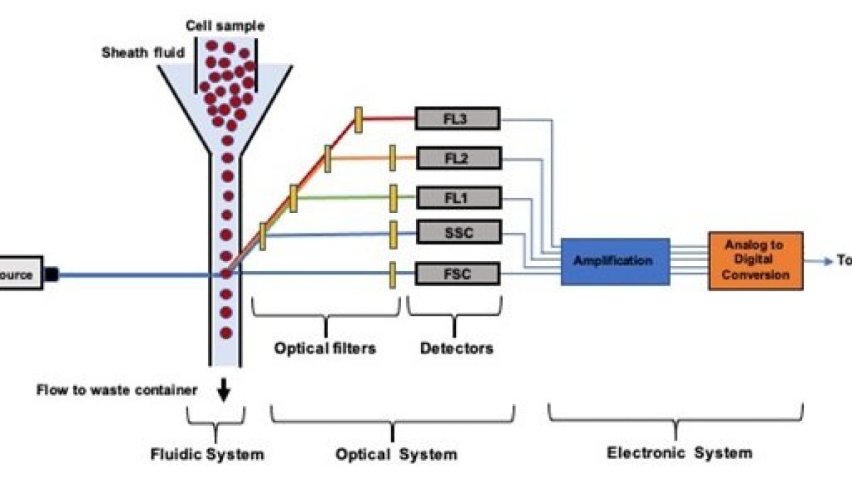
Our high-performing flow cytometers equip you for a broad of range of clinical and research applications including measurement of CD4 counts in HIV patients residual white blood cells in blood transfusions CD34 hematopoietic cells for stem cell transplantation and many more basic and clinical research targets. Flow cytometry was performed on a BD FACScan flowcytometry system. If cells are to be collected for.
Centrifuge at 1200-1500 rpm for 5 minutes. Harvest wash the cells and adjust cell suspension to a concentration of 1-5 x 10 6 cellsmL in ice-cold PBS 10 FCS 1 sodium azide. This incubation must be done in the dark.
Either BD 352350 sterile cell strainer cap fits 50ml tube or BD Falcon 352235. The purpose of the azide in these buffers is to prevent microbial growth but these buffers are used so quickly and are extremely cheap to make that you shouldnt run into any problems. However they can be stained in any container for which you have an.
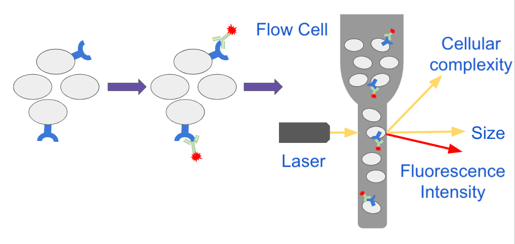
Flow cytometry FACS staining protocol Cell surface staining 1. Flow cytometry is a popular cell biology technique that utilizes laser-based technology to count sort and profile cells in a heterogeneous fluid mixture. Store the cell suspension immediately at 4C in the dark.
FACS Staining Buffer 1XPBS w 3 calf serum and 005 sodium azide Collection Buffer depends on application RPMI or PBSserum 70uM filter. Flow cytometry and FACS fluorescence activated cell sorting are distinctly different procedures though FACS is a descendant procedure based upon flow cytometry. If cells are to be collected for.
Prepare single-cell suspensions from either lymphoid tissue bone marrow peripheral blood or cell cultures using standard protocols. FACS Buffer - Mar132012 FACS Buffer -. Add 1 ml PBS to rinse non-bound antibody.
Here are 5 ingredients to consider for your FACS buffer. Using FACS a researcher can physically sort a heterogeneous mixture of cells into different populations. 424401 True-Nuclear Transcription Factor Buffer Set.
Cell Surface Staining of Human PBMCs and Cell Lines. This Flow Cytometry Staining Buffer is a buffered saline solution containing fetal bovine serum and sodium azide 009 as a preservative. This buffer can be used for antibody and cell dilution steps as well as all the wash steps required for the surface staining and flow cytometric analysis.
Wash the cells 3 times by centrifugation at 400 g for 5 min and resuspend them in ice-cold PBS 3 BSA 1 sodium azide. 1 Phosphate Buffer Saline or Hanks Buffer CaMg2 free 1 mM EDTA 25 mM HEPES pH 70 05-2 Fetal Bovine Serum Heat-inactivated or 1 BSA 02 μm sterile filtered Store at 4C For Clean Lymphoid Cells. By using highly specific antibodies tagged with fluorescent dyes a researcher can perform FACS analysis and simultaneously gather data on and sort a sample by a nearly limitless number of.
We use this buffer. Prepare the following buffer in which to suspend cellular samples prior to cell sorting. Harvest wash the cells single cell suspension and adjust cell number to a concentration of 1-5106 cellsml in ice cold FACS Buffer PBS 05-1 BSA or 5-10 FBS 01 NaN3 sodium azide.
1- Use CaMg2 free PBS. Weve had a basic introduction to flow cytometry and the machine is running relatively well however we frequently have problems with clogs. General procedure for flow cytometry using a conjugated primary antibody.
Primary Antibody Staining 1. Add 1 μg of primary antibody directly to 50-100 μl of suspended cells. This buffer can be used for antibody and cell.
554656 Stain Buffer FBS 420201 Cell Staining Buffer. Use of FCS or BSA in in FACS buffer reduces autofluorescemce caused by non specific biding by antibodies which may falsely increase the MFI of a channel in flow cytometery Cite 2 Recommendations. Flow cytometry FACS staining protocol Cell surface staining Harvest wash the cells single cell suspension and adjust cell number to a concentration of 1-5x10⁶ cellsml in ice cold FACS Buffer PBS 05-1 BSA or 5-10 FBS 01 NaN3 sodium azide.
Incubate for at least 20-30 min at room temperature of 4C. Another reason that people use protein containing buffers for flow cytometry is to prevent cells from sticking to the side of plastic tubes or other culture labware as well as preventing cell. Flow Cytometry Direct immunofluorescence staining.
Add 1 mL FACS Buffer to each tube. Incubate on ice for 20 minutes. Im a relatively new FACS user as is everyone in my lab.
FACS is an abbreviation for fluorescence-activated cell sorting which is a flow cytometry technique that further adds a degree of functionality. We use this buffer for surface staining as well as for intracellular staining. FACS is a derivative of flow cytometry that adds an exceptional degree of functionality.
A debris free single cell suspension will have lower auto-fluorescence and flow smoothly through the. The additional cations in the recipe promote better viability. Staining buffer is the buffer used during.
If the nozzle isnt clogged then there is something blocking or at least causing problems with the. Absence of these ions reduces cation-dependent cell to cell adhesion and prevents clumping. Basic Sorting Buffer 1 x Phosphate Buffered Saline PBS or Hanks Balanced Salt Solution HBSS Ca2 Mg2 Free 1mM EDTA 25 mM HEPES pH70 1 Fetal Calf Serum Heat inactivated or 1 Albumin - Filter sterilize using a 02 µM filter - Store at 4 degrees.
Add 100 μl of the cell suspension to each tube.

Key Steps In Flow Cytometry Protocols
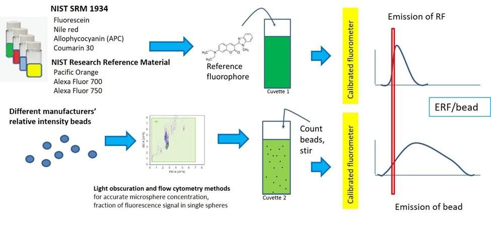
Quantitative Flow Cytometry Measurements Nist
Facs Buffer Composition

Facs Buffer Composition
Flow Cytometry Perm Buffer 10x Pf00011 C Proteintech

We Offer Anti Mouse Cd3 Antibody Biot Conjugated Keep As Concentrated Solution Store At 4 C And Protected From Prolonged Exposure To Life Science Index Biot
Ebioscience Flow Cytometry Staining Buffer

Fundamentals Of Flow Cytometry Aat Bioquest
Microfluidic Flow Cytometry Principles And Commercial Review Ufluidix

Flow Cytometry And Cell Sorting By Facs In The Flow Cell 1 The Download Scientific Diagram
Facs Buffer Composition

What Is Flow Cytometry Technology Networks
Popular Antibodies For Flow Cytometry Proteintech Group
Flow Cytometry Cell Analysis Vs Cell Sorting Products Bio Rad
Facs Buffer Composition

Ep2 Polyclonal Antibody Calculated Mw 40 Kda Observed Mw 40 Kda Source Rabbit Isotype Igg For More Information Click On Life Science Observation Index
Analyzing Single Cells With Flow Cytometry
Facs Buffer Composition
Flow Cytometry And Cell Sorting By Facs In The Flow Cell 1 The Download Scientific Diagram